Following the previous success, the Environmental Mineralogy Group (EMG) is once again launching its photo competition!
This time, we plan to showcase the best environmental mineralogy has to offer across not only EMG’s publications and website, but also at the forthcoming Mineralogical Society’s 150th Anniversary conference: https://minsoc-150.org/
If you aren’t familiar with the competition, the competition is open to all areas of environmental mineralogy and submission is easy. All that’s required is for you to submit your favourite image, be that out in the field, in the laboratory, or even under the (electron) microscope). Accompany your image with a description about what’s going on in your image and tell everyone why what you’ve seen is so interesting.
Submitting to the competition is easy, simply send your entry to the email address below with a description of your photo detailing why your entry is interesting. We welcome entries for all stages of careers in environmental mineralogy; it doesn’t matter if you’re a student or an established expert, we want to see your photos!
This year, all entries will be showcased at the Mineralogical Society’s 150th Anniversary conference throughout the 3-day event – including at the EMG stand and on the big screens before and after talks. Additionally, and the winning photos will be displayed on the EMG website, EMG / MinSoc Newsletters, and published in Elements magazine. Finally, the best submission will receive a monetary prize following judging by the EMG committee.
The deadline for submissions is Friday 15th May 2026.
Submit your entries (or any queries you may have) here: emg@minersoc.org
Here are a couple of examples to get you started:
Example 1:
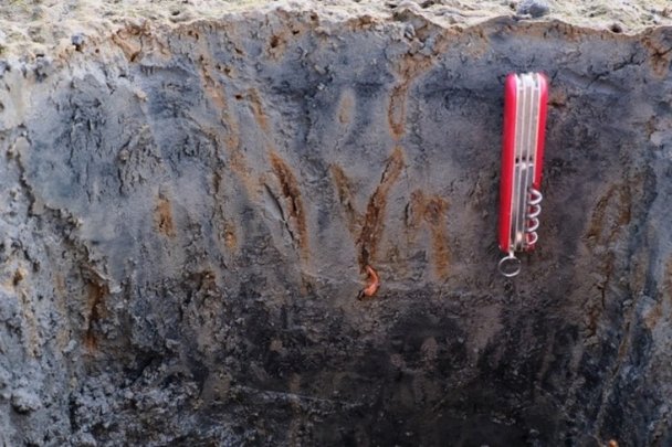
A profile of intertidal flat sediments in the German Wadden Sea. The image shows several redox features including the precipitation of iron (oxyhydr)oxides (orange/brown) in lugworm burrows formed as a result of oxygen in the burrows oxidising reduced iron in the otherwise grey sediment. The sediment becomes darker/black with depth indicating the formation of a redox profile.
The sediments likely undergo cycles of reduction during tidal flooding and oxidation during the ebb tide, causing the iron minerals in the sediment to dynamically precipitate and dissolve. We are researching the implications of these iron mineral transformations for the cycles of nutrients and contaminants.
Example 2:
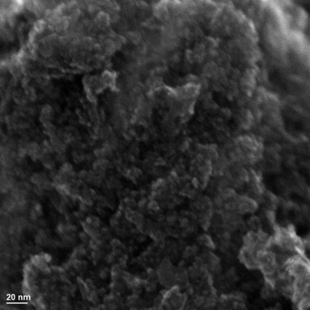
SEM image of ferrihydrite. While this image may look unimpressive with no discernible crystalline structures visible at the nanometre scale, this highlights the importance of ferrihydrite (a short range ordered iron oxyhydroxide) in the environment. Due to its short range ordered nature, ferrihydrite has an extremely high specific surface area and is therefore an important sorbent for contaminants and nutrients.
Last Year’s Winner (Brooke Johnson)
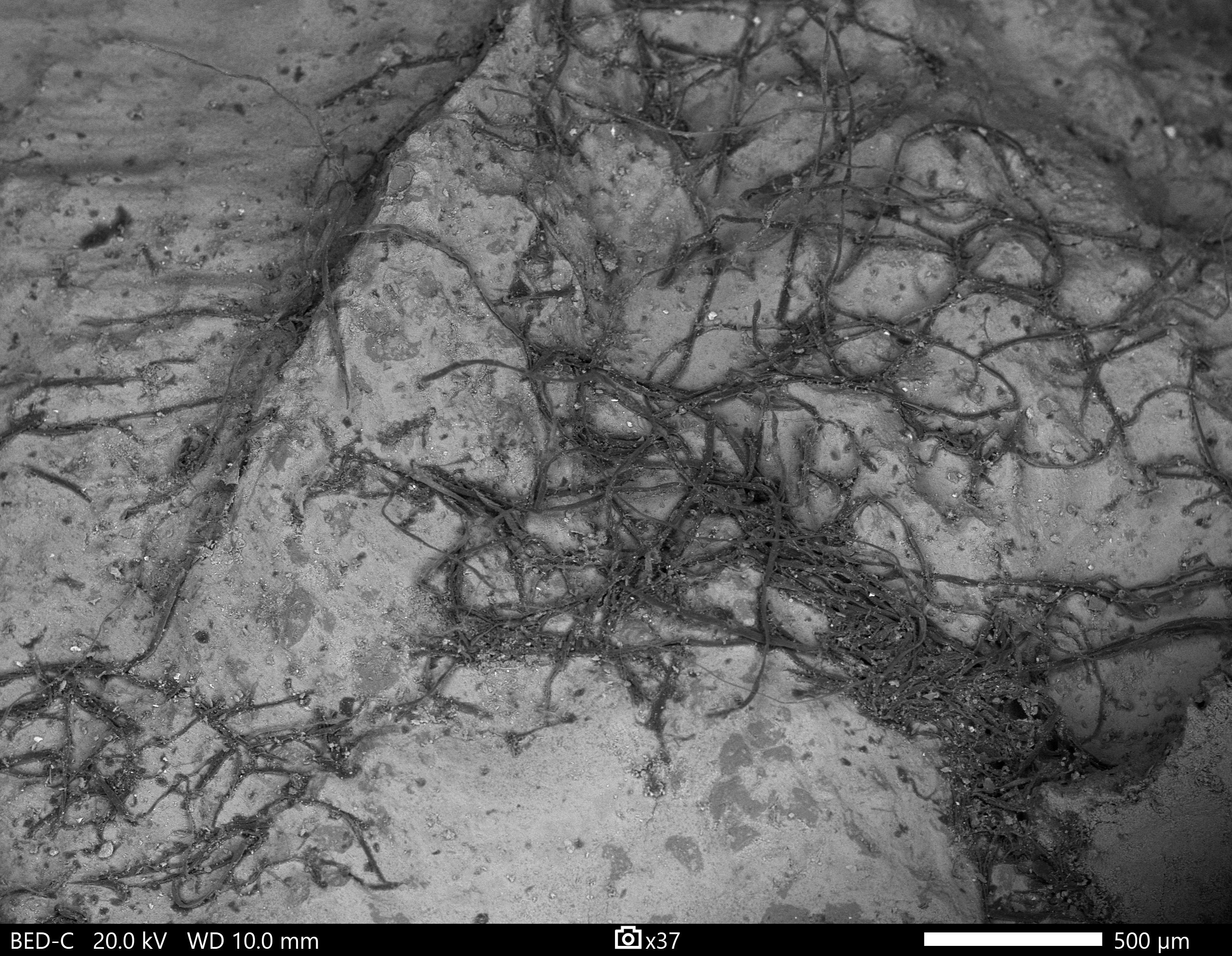
The entry is a backscatter electron image of the exposed internal cortex surface of a 1.4 billion year old Fe-phyllosilicate ooid. The black filaments are exceptionally preserved 3-dimensional fossil acritarchs that were entrained within the cortex of the grain during deposition. The pale grey matrix is interlaminated berthierine and greenalite, the mid grey grains are silica with features suggesting they maybe silicified acritarchs. The latter point is supported by the observation that the organic filaments all host a silica internal fill. This unit contains abundant exceptionally preserved acritarchs which retain nanoscopic structural features not previously observed in fossils of this age. I hypothesise that rapid Fe-phyllosilicate authigenesis in a hydrodynamically active depositional setting contributed to the exceptional preservation of these acritarchs. This finding is also significant because Precambrian ferruginous sedimentary rocks are canonically barren of fossils and low in organic matter. Our data suggest that examining the unoxidised green clay facies of these units may provide new window into Precambrian life.